The Cell and Molecular Biology Graduate Program is an interdisciplinary program comprised of over 130 faculty from three colleges. Research interests of faculty on the CMB Graduate Studies Committee are quite varied allowing the program to offer focused study in any of seven research tracks:
Click on a research Area below to see the faculty.
Bioinformatics & Computational Biology
Comprised of an interdisciplinary and collaborative group of researchers who are interested in topics that span all aspects of biology, biochemistry, biophysics and neurobiology. A distinguishing characteristic of the students in this track, in contrast with the students in the other Cell and Molecular Biology tracks, is the magnitude and sophistication of the computational component of their project. This includes but is not limited to - the analysis of large data sets, more exhaustive computational analysis, develop novel types of analysis, and write sophisticated computer programs to analyze their data. Interactions and collaborations with investigators from other CMB tracks, Biostatistics, and the departments of Computer Science, Engineering, and Biomedical Engineering are encouraged. Students who do well in this track frequently demonstrate individual initiative in generating and executing projects. Students are allowed to have more than one CMB mentor during their graduate careers to develop and implement interdisciplinary projects.
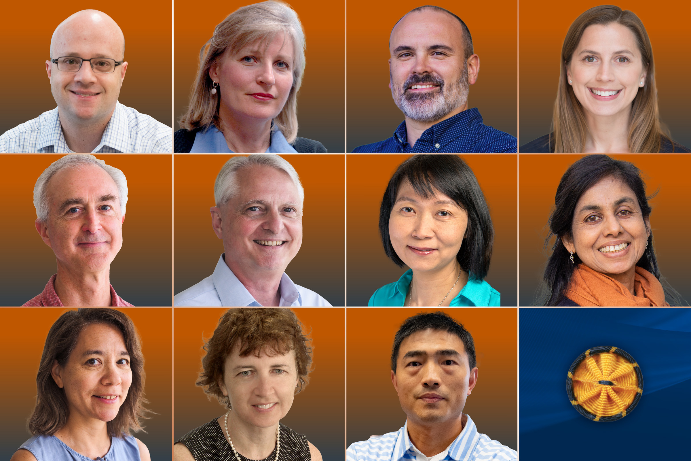
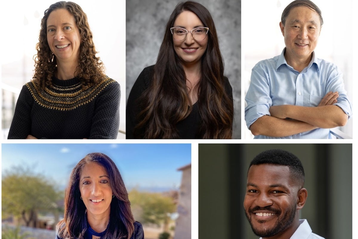
Faculty:
Alper, Hal
Anslyn, Eric
Barrick, Jeffrey
Bayer-Santos, Ethel
Brock, Amy
Bull, James
Cenik, Can
Cenik, Elif
Contreras, Lydia
Elber, Ron
Ellington, Andrew
Georgiou, George
Gutell, Robin
Havird, Justin
Hillis, David
Hofman, Hans
Jansen, Bob
Lu, Yi
Makarov, Dmitrii
Marcotte, Edward
Markey, Mia
Meyers, Lauren
Miranker, Daniel
Press, William
Taillefumier, Thibaud
Wang, Huilang (Evan)
Wilke, Claus
Yi, Stephen
Zhang, Yan (Jessie)
Biomolecular Structure & Function
Research on how proteins and nucleic acids are organized, and their structure and function in cells. Using powerful techniques such as X-ray crystallography or NMR spectroscopy, as well as biochemical or biophysical methods, models of macromolecules and their properties are used to explain the molecular basis of catalysis, recognition and disease.
Faculty
Appling, Dean
Bajaj, Chandrajit
Barrick, Jeffrey
Bayer-Santos, Ethel
Belardi, Brian
Browning, Karen
Cambronne, Lulu
Cenik, Can
Contreras, Lydia
Dalby, Kevin
Dickinson, Daniel
Eberlin, Livia
Elber, Ron
Ellington, Andrew
Fast, Walter
Finkelstein, Ilya
Georgiou, George
Hackert, Marvin
Hoffman, David
Jara Oseguera, Andres
Johnson, Kenneth
Keatinge-Clay, Adrian
Lambowitz, Alan
Leahy, Dan
Liu, Ben
Lu, Yi
Marcotte, Edward
Matouschek, Andreas
Maynard, Jennifer
McLellan, Jason
Mukhopadhyay, Somshuvra
Newberry, Robert
Robertus, Jon
Russel, Rick
Taylor, David
Webb, Lauren
Whitman, Christian
Yi, Stephen
Zhang, Jessie
Chemical Biology & Drug Discovery
Research at the chemistry-biology interface that focuses on the discovery and development of drugs and health-related technology. The faculty members have extensive experience in the synthesis of organic and biopolymer therapeutics, the development of high-throughput screens and assays, the application of three dimensional structure determination and modeling techniques to drug design, and the identification and evaluation of molecular targets and therapeutics, and the evaluation of the toxic action of drugs and drug candidates.
Faculty
Belardi, Brian
Cambronne, Lulu
Dalby, Kevin
Eckhardt, Gail
Ellington, Andrew
Fast, Walter
Johnson, Kenneth
Keitz, Keith Benjamin
Lee, Seongmin
Liu, Hung-Wen
Lu, Yi
Marcotte, Edward
Martin, Stephen
Maynard, Jennifer
Mills, Edward
Ren, Pengyu
Richburg, John
Torii, Keiko
Tiziani, Stefano
Umlauf, Ben
Whitman, Christian
Yi, Stephen
Zhang, Yan Jessie
Cell & Developmental Biology
Research on the cell biology and development of animal, plant and microbial systems. Faculty affiliated with this track have a broad diversity of research interests including topics such as signal transduction, membrane traffic, cell polarity and motility, regulation of gene expression, cell-cell interactions, specification of cell identity, spatial patterning of developing embryos and evolution of developmental mechanisms. There is also a strong commitment to imaging techniques including scanning laser confocal microscopy, in vivo imaging of developmental processes, measurements of intracellular ion concentrations and electron microscopy.
Faculty
Baker, Aaron
Belardi, Brian
Brock, Amy
Cambronne, Lulu
Cenik, Can
Cenik, Elif
Crews, David
De Lozanne, Arturo
Dickinson, Daniel
DiGiovanni, John
Eberhart, Johann
Ellington, Andrew
Finnell, Richard
Fischer, Janice
Florin, Ernst-Ludwig
Gordon, Vernita
Gray, Ryan
Gross, Jeffrey
Huq, Enamul
Iverson, Brent
Jolly, Christopher
Juenger, Thomas
Keatinge-Clay, Adrian
Kim, Jonghwan
Kuo, John
Lloyd, Alan
Lu, Yi
Macdonald, Paul
Marcotte, Edward
Matouschek, Andreas
Mills, Edward
Mukhopadhyay, Somshuvra
O'Halloran, Theresa
Poenie, Martin
Qiao, Hong
Roux, Stanley
Senning, Eric
Stachowiack, Jeanne
Stein, David
Suggs, Laura
Sung, Sibum
Torii, Keiko
Van Den Berg, Carla
Vokes, Steven
Wallingford, John
Woznica, Arielle
Yi, Stephen
Molecular Genetics
Fundamental questions are asked concerning inheritance and changes of genetic information in living organisms. In molecular terms, this information is encoded at the level of DNA, RNA, and/or protein. The track includes diverse fields of biology ranging from cellular processes including DNA recombination, repair, transcription, splicing, translation, genomics, gene silencing, chromatin modifications, epigenetic mechanisms, RNA interference, microRNA regulation, and control of the cell-division cycle, to molecular evolution as well as human cancers and other diseases. Faculty affiliated with this track represent a broad range of research interests and experimental systems from phage, virus, and bacteria to yeast, plants, flies to mice and humans.
Faculty
Alper, Hal
Barrick, Jeffrey
Bayer-Santos, Ethel
Cambronne, Lulu
Cenik, Can
Cenik, Elif
Chen, Zengjian (Jeff)
Croyle, Maria
Davies, Bryan
Dudley, Jaquelin
Ehrlich, Lauren
Ellington, Andrew
Georgiou, George
Gross, Jeffrey
Harshey, Rasika
Huibregtse, Jon
Iyer, Vishwanath
Jayaram, Makkuni
Jiang, Ning
Johnson, Arlen
Keitz, Keith Benjamin
Lambowitz, Alan
Lu, Yi
Marcotte, Edward
Matouschek, Andreas
Matz, Mikhail
Meyer, Richard
Miller, Kyle
Molineux, Ian
Moran, Nancy
Ochman, Howard
Paull, Tanya
Payne, Shelley
Stevens, Scott
Sullivan, Christopher
Torii, Keiko
Tucker, Haley
Upton, Jason
Vasquez, Karen
Walker, James
Woznica, Arielle
Xhemalace, Blerta
Yi, Stephen
Neurobiology
Primary interests include applying cellular and molecular techniques to study the nervous system. Our faculty have wide ranging research interests that touch on most of the present frontiers in neuroscience, including specification of neuronal cell fates, integration of information by synapses and dendrites, alterations in neuronal circuits as a consequence of experience, drug and alcohol addiction, sensory processing, transmitter release, repair of neuronal injuries, hormones and behavior, learning and memory, and gene expression.
Faculty
Agarwala, Seema
Aldrich, Richard
Atkinson, Nigel
Ben-Yakar, Adela
Bittner, George|
Cambronne, Lulu
Champagne, Frances
Drew, Michael
Fonken, Laura
Ellington, Andrew
Golding, Nace
Gore, Andrea
Gross, Jeffrey
Harris, Adron
Hofman, Hans
Lee, Amy
Lu, Yi
Marcotte, Edward
Matsui, Bill
Messing, Robert
Mihic, John
Morikawa, Hitoshi
Nishiyama, Hiroshi
Phelps, Steven
Pierce, Jonathan
Priebe, Nicholas
Shear, Jason
Wang, Evan
Zakon, Harold
Zemelman, Boris
Plant Molecular Biology
The Plant Molecular Biology Track brings diverse faculty together that use plants or photosynthetic organisms as model systems for the study of basic biological processes, genomics, and biofuels.
Faculty
Browning, Karen
Chen, Jeff
Ellington, Andrew
Hawkes, Christine
Huq, Enamel
Jansen, Robert
Juenger, Thomas
Lloyd, Alan
Lu, Yi
Marcotte, Edward
Mehdy, Mona
Moran, Nancy
Qiao, Hong
Roux, Stanley
Sung, Sibum
Torii, Keiko